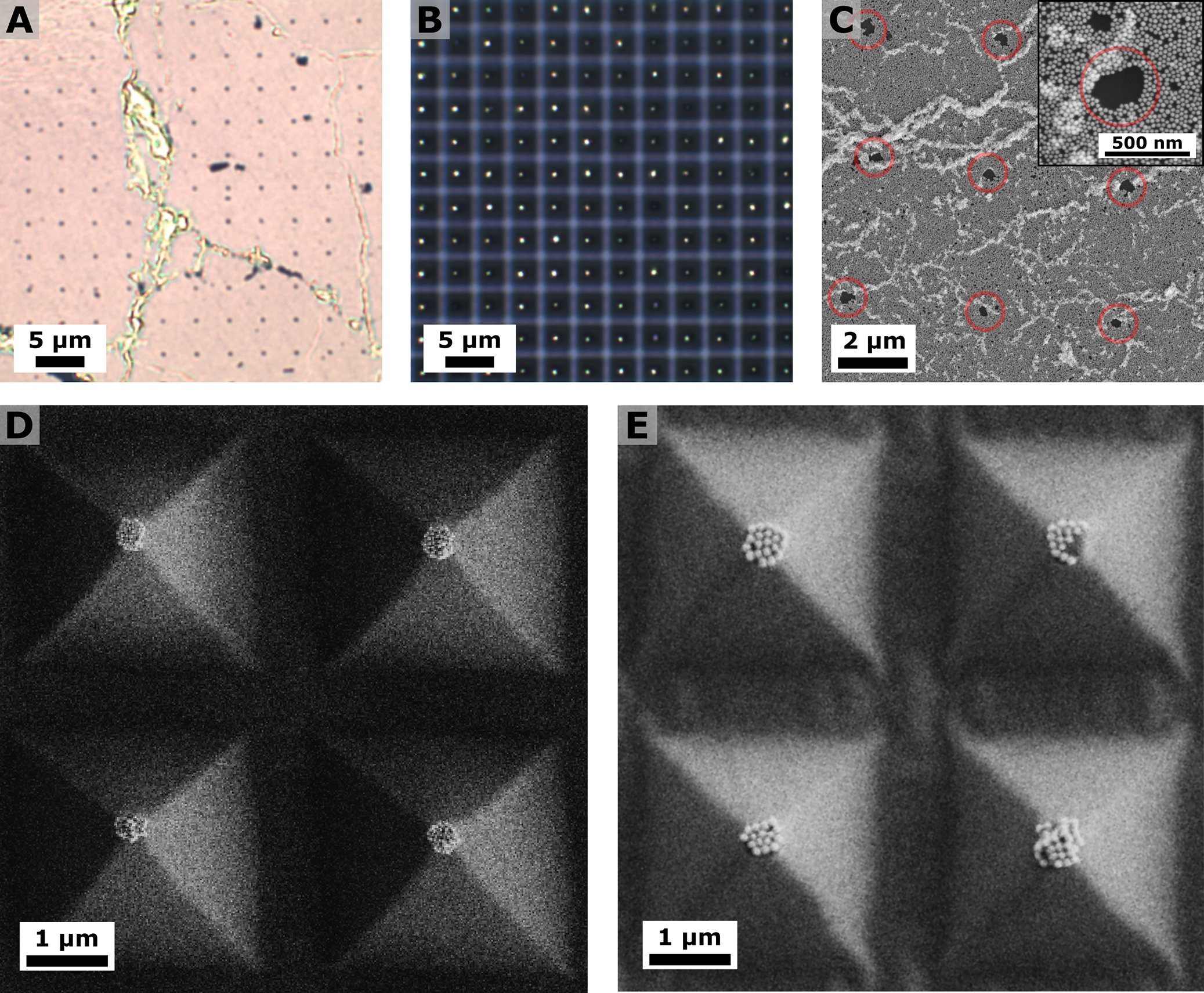
Figure 4:

(A,B) Bright-field micrographs of the complementary negative image with the imprinted pyramid pattern into the nanoparticle monolayer (A) and the patterned polydimethylsiloxane (PDMS) after lift-off (B). (C) Scanning electron microscopy (SEM) analysis of the complementary negative image. Inset: higher magnification image of one circled area. (D-F) SEM of the patterned PDMS after lift-off using different nanoparticle dimensions: 50 ± 3 nm (D), and 100 ± 9 nm (E). SEM images of the patterned PDMS after lift-off applying low pressures are provided in Figure S9.
