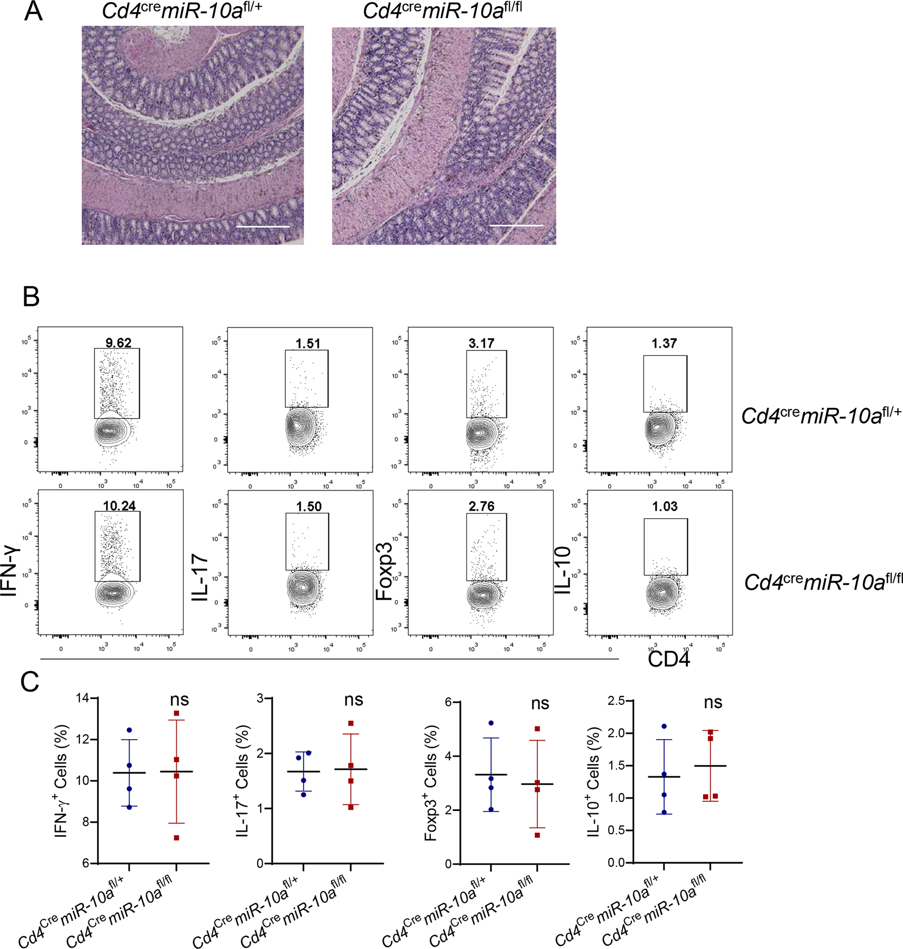
Figure 1. Deficiency of miR-10a in CD4+ T cells does not affect intestinal CD4+ T cell differentiation in vivo under steady conditions.

10-week age-matched wild type (WT) Cd4cremiR-10afl/+ and CD4 T cell-specific miR-10a deficient Cd4cremiR-10afl/fl mice were sacrificed. (A) Representative H&E staining of colon was shown. Scale bars, 300 μm. (B-C) Flow cytometry profile (B) and Quantification (C) of IFN-γ +, IL-17+, Foxp3+, and IL-10+ CD4+ T cells in intestinal lamina propria were shown. All data are presented as mean ± SD. One representative of two independent experiments. Unpaired Student’s t test; ns, not significant.
