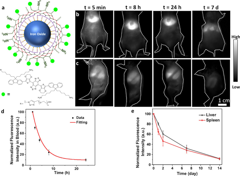
Figure 7.
Excretion of iron oxide nanoparticles with P3 coating. a) Schematic illustration of iron oxide nanoparticles with P3 coating and conjugated to IR-FEPC. Wide-field NIR-II (1000–1200 nm) fluorescence images of mice showed decay of the fluorescence signal over time in b) liver and c) spleen. The mice were excited by an 808 nm laser at a power density of 70 mWcm2. d) NIR-II fluorescence signal in femoral artery of the mice at different time points post injection. The iron oxide nanoparticles showed a blood circulation half time of 2.86 hours. e) Fluorescence signal decay of liver and spleen over the course of 2 weeks.

