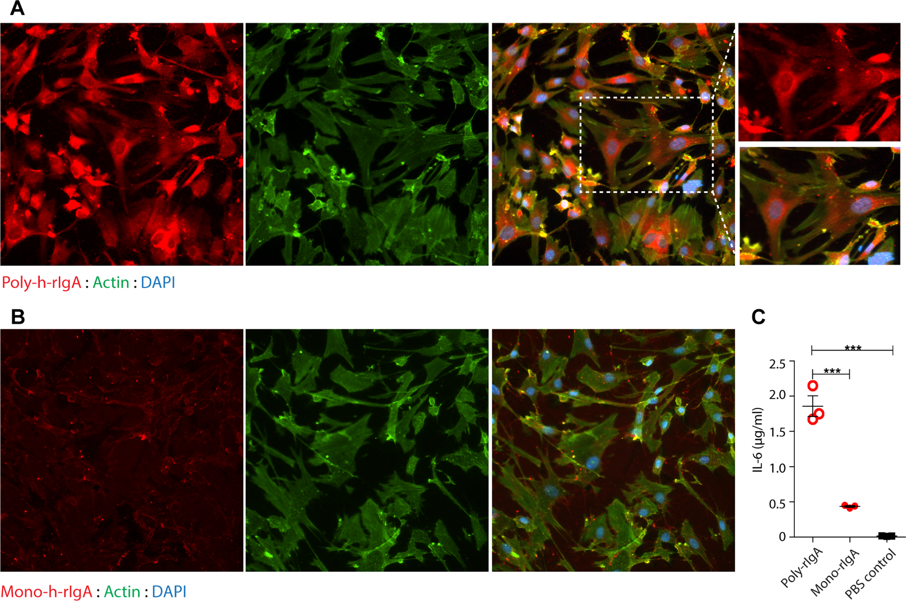
Figure 6.

Poly-IgA binds renal mesangial cells in culture. (A, B) Human glomerular mesangial cells were cultured in dishes. Human biotin-h-rIgA either in the SA-induced polymeric state (A) or in the uninduced monomeric state (B) was added to the culture medium. Following washing to remove unbound h-rIgA, the cells were fixed and then probed for IgA contents. Phalloidin staining for actin and DAPI for DNA were used as counterstains. While strong poly-h-rIgA signals were associated with the cells (in A), there was no specific binding of mono-h-rIgA detected (in B). (C) Following co-cultureofthecells with either poly- or mono-h-rIgAovernight, theculture mediumwasharvestedfor detectionof IL-6 by ELISA (y-axis). The inflammatory response of the cells (IL-6 production) to human poly-hrIgA was significantly greater than that to mono-h-rIgA.
