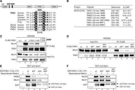
Fig. 2. An adjacent MLH1-interacting motif (MIM) in FAN1 synergizes with the MIP box to promote stable MLH1 binding.

(A) Top: Protein domain architecture of human FAN1 indicating the location of the MIP box and MIM. Bottom: Sequence alignment of FAN1 orthologs with the putative MIM of human MLH3, PMS2, and PMS1. (B) ITC was used to determine the Kd indicating the binding affinity of MLH1-CTD for the indicated peptides. (C) Immunoblots of FLAG-M2 affinity resin IPs of extracts from HEK293 cells transfected either with e.v. or indicated FLAG-FAN1 expression constructs. The L155A/L159A mutation is denoted as MIM*. (D) Immunoblots of FLAG-M2 affinity resin IPs of extracts from HEK293 cells transfected either with e.v. or indicated FLAG-FAN1 expression constructs. (E and F) Recombinant MutLα (E) or MutLγ (F) (200 nM) was subjected to pull-down reactions using the indicated recombinant GST-FAN1 (amino acids 118 to 177) variants. (C to F) The antibodies used are shown on the left.
