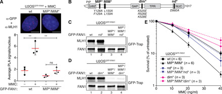
Fig. 3. FAN1-MLH1 interaction promotes resistance to cross-link damage.

(A) PLA was used to evaluate FAN1-MLH1 association in U2OSGFP-FAN1 wt and U2OSGFP-FAN1 MIP*/MIM* cells mock-treated or treated with MMC (150 ng/ml) for 24 hours. Representative images are shown. Scale bars, 10 μm. Scatterplot displays quantification of the PLA signals per nucleus from at least 100 cells. Data display the means ± SD from three independent experiments. Statistical significance was calculated by unpaired t test. **P < 0.01; ns, not significant. (B) Schematic representation of human FAN1, highlighting positions of mutations used, including MIP*, MIM*, dimerization-defective (dim*), and nuclease-defective (nd*) FAN1 variants. (C and D) Immunoblots of GFP-Trap IPs of extracts from indicated U2OSGFP-FAN1 cells. The antibodies used are shown on the left. (E) Clonogenic survival assay of the indicated U2OSGFP-FAN1 cells exposed to increasing doses of MMC. Viability of untreated cells was defined as 100%. Data are presented as the means ± SEM.
