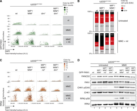
Fig. 4. FAN1-MLH1 interaction prevents extensive ssDNA formation following cross-link damage.

(A) QIBC analysis of GFP-FAN1 foci in U2OSGFP-FAN1 cells mock-treated, treated with MMC (20 ng/ml) for 24 hours, or MMC-treated and then released for 72 hours. Color-coded scatterplots indicate the number of GFP-FAN1 foci per nucleus. ut, untreated. (B) Same cells as in (A) were pulse-labeled with ethynyl deoxyuridine (EdU) during the last 30 min before harvesting and subjected to the Click-IT reaction. Cell cycle distribution was evaluated by QIBC using the 4′,6-diamidino-2-phenylindole (DAPI) and EdU signals (fig. S5C). (C) QIBC of RPA2 foci in U2OSGFP-FAN1 cells mock-treated, treated with MMC (20 ng/ml) for 24 hours, or MMC-treated and then released for 72 hours. Color-coded scatterplots indicate the number of RPA2 foci per nucleus. A.U., arbitrary units. (D) Same cells as in (C) were treated with MMC (300 ng/ml) for 24 hours, and lysates were analyzed by immunoblotting using the indicated antibodies. Asterisk indicates hyperphosphorylated form of RPA2.
