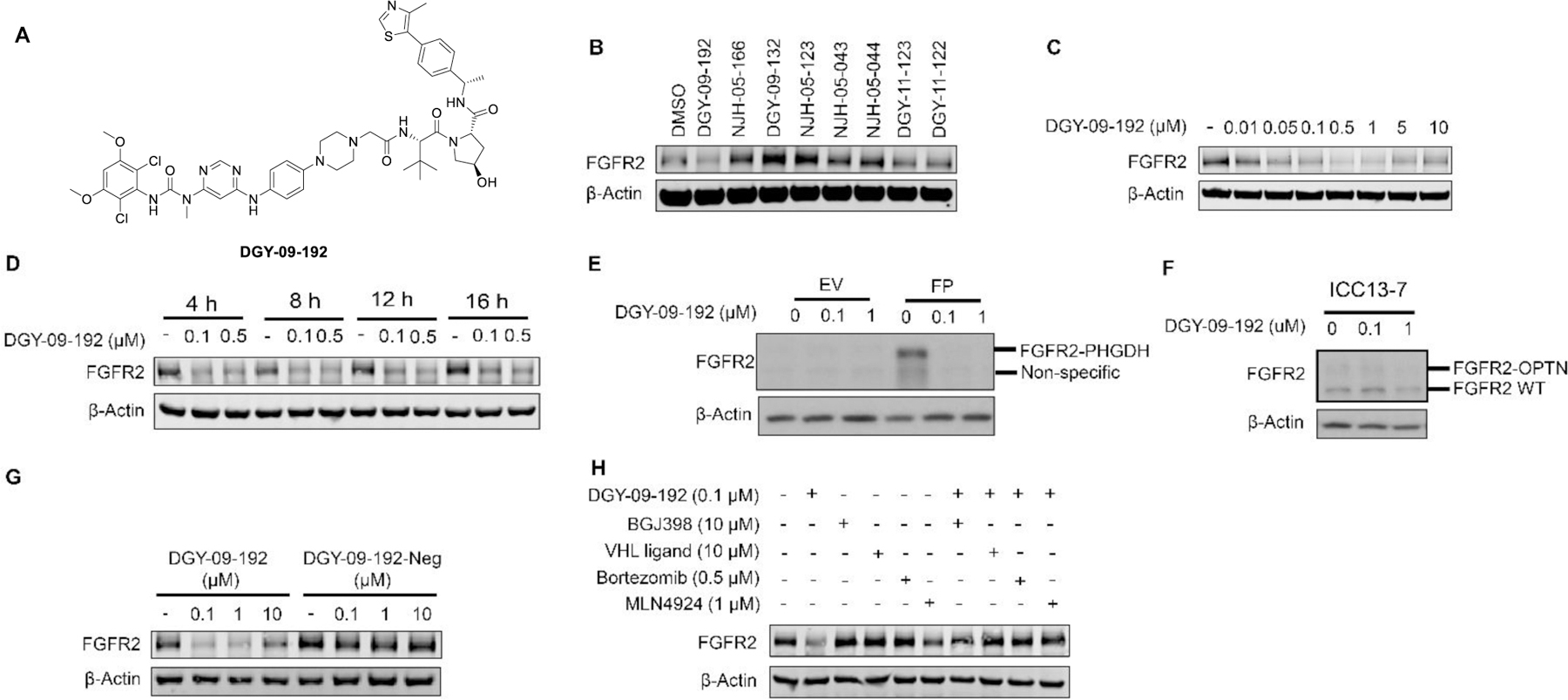
Figure 3.

DGY-09-192 induced degradation of FGFR2 or FGFR2-fusion in a VHL dependent manner. A. Structure of DGY-09-192. B. Immunoblot analysis of FGFR2 in Kato III cells treated with 1 µM of the indicated compounds for 24h. C. Immunoblot analysis of FGFR2 in Kato III cells treated with indicated concentrations of DGY-09-192 for 6h. D. Immunoblot analysis of FGFR2 in Kato III cells treated with DGY-09-192 at indicated time points. E. Immunoblot analysis of FGFR2-PHGDH (FP) fusion in CCLP-1 cells and F) FGFR2-OPTN fusion in ICC13–7 cells treated with DGY-09-192 for 24h. G. Immunoblot analysis of FGFR2 in Kato III cells treated with DGY-09-192 or DGY-09-192-Neg for 4h. H. Immunoblot analysis of FGFR2 in Kato III cells pretreated for 2h with BGJ398, VHL ligand, Bortezomib or MLN4924, and then treated with DGY-09-192 for 4h. EV is short for empty vector.
