
FIG 1.
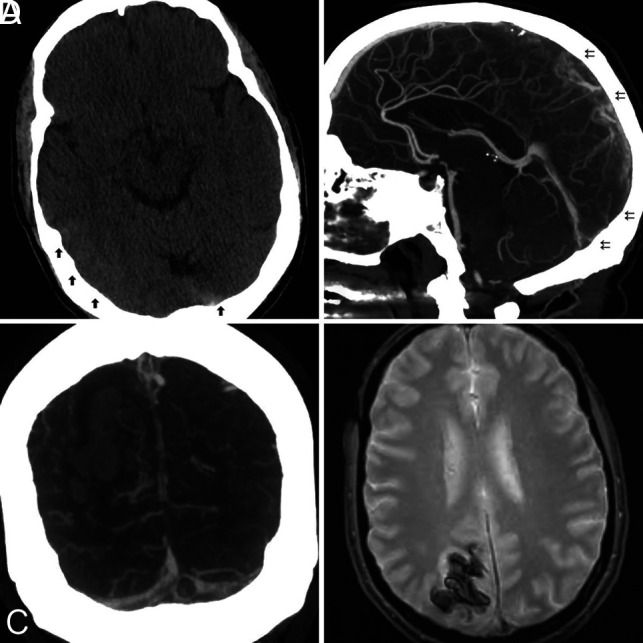
A, NCCT shows hyperdense thrombus in the region of the torcula and subtle cortical hyperdensity along the right temporo-occipital region (arrows). Sagittal (B) and coronal MIP (C) reformatted images of CTA show multiple filling defects along the posterior superior sagittal sinus (double arrows), torcula, and right transverse sinus. Note a parenchymal hematoma in the right occipital lobe. D, MR imaging of the brain; gradient recalled-echo sequence shows thrombus in the sagittal sinus and associated parenchymal hematoma.