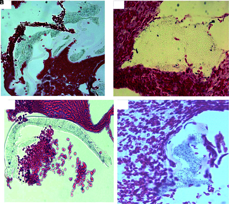
FIG 3.
Representative microphotographs of scratched pieces of different layers rather than the surface or inner layer. Microphotographs showing the different materials of MT catheter, rather than the surface-coating material and inner liner materials shown in Figs 1 and 2. The similar foreign material is found in the patients’ retrieved clot tissue (type III, Fig 7) (H&E, original magnification ×400).

