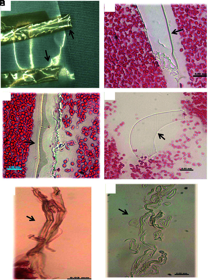
FIG 4.
Histologic features of donated PTFE material. A, Macrophotograph of the PTFE liner material provided by manufacturer sponsor. B–F, Microphotographs of the scratched PTFE pieces in H&E–stained slides. The material appears to have varied shape. It is either a mass with solid, homogeneous texture associated with refraction, and light green edge (B–D), or long strips or tubes with light green outline (E and F). Those features are the same as the scratched inner layer of catheters (Fig 2), and type I material found in the patient clot tissue as well (Fig 5) (B–F, H&E, original magnification ×400).

