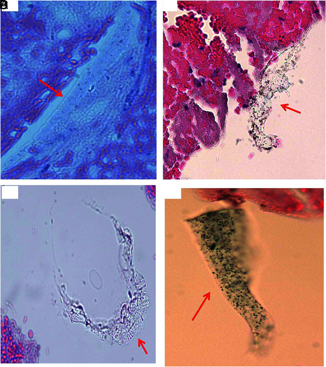
FIG 7.
Representative microphotographs of type III foreign material found in patient clot tissue. A, A single, long, oval-like piece of foreign material embedded within clot tissue (arrow). The material is associated with multiple bubblelike particles, which are refracted under light microscopy. B, A single piece of irregular foreign material attaching to the clot tissue (arrow). It appears to be light green, associated dark/black and bubblelike, with refraction particles. C, Irregular piece of foreign material at the gap of clot tissue (arrow). It appears to be solid, homogeneous in texture with refraction, and light green. D, A piece of green, solid foreign material, associated diffused black, dotlike particles (arrow) (A–D, H&E, original magnification ×400). These materials presented in A–D are similar to the materials scratched from the catheters (Fig 3).

