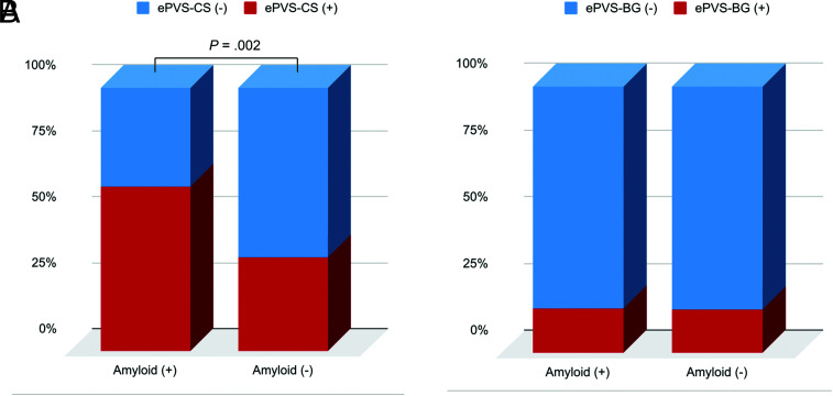
FIG 2.
Comparisons of the presence of MR imaging–visible PVS-CS (A) and MR imaging–visible PVS in the basal ganglia (B) based on the β-amyloid status. The enlarged perivascular spaces in the centrum semiovale (ePVS-CS) were significantly higher in the patient group positive for β-amyloid than in the patient group negative for it, whereas the high degree of enlarged perivascular spaces in the basal ganglia (ePVS-BG) did not differ between the groups positive and negative for β-amyloid.

