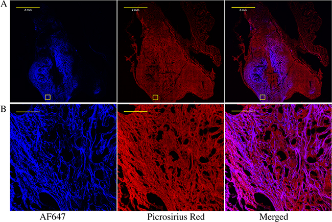
Figure 3.
Imaging NHS-ester and extracellular matrix co-localization within a pancreatic tumor. (A) Whole tumor and (B) zoom-in images of the boxed area of fluorophore-NHS-injected tumors stained for extracellular proteins with picrosirius red. Pancreatic KPC 4662 tumors were injected intratumorally with AF647 NHS ester. After 24 h, tumors were excised, fixed, sectioned, and stained with picrosirius red. The scale bars for (A) and (B) are 2 mm and 100 μm, respectively.

