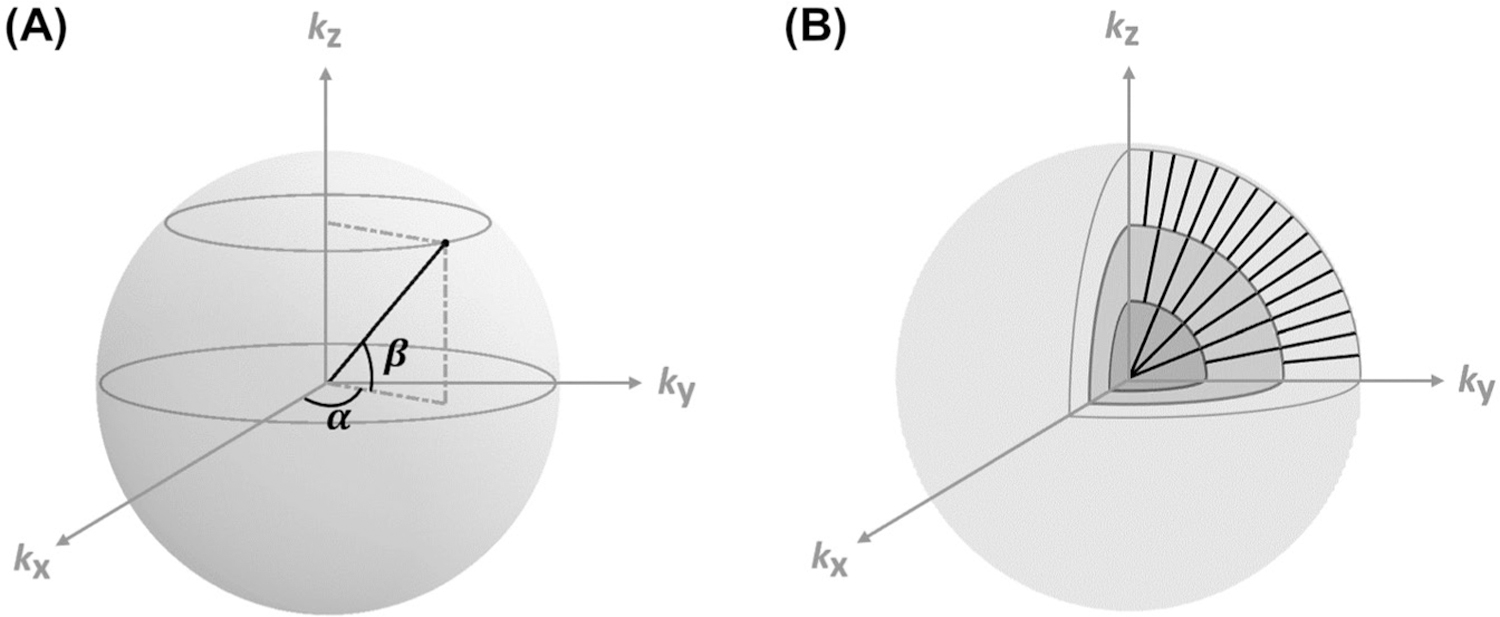
FIGURE 1.

Schematics of data sampling and image reconstruction. (A) Golden-means-based radial sampling. α and β are the azimuthal and polar angles of a radial spoke, respectively. (B) KWIC-based image reconstruction. Only 3 concentric shells in the partitioned k-space are shown. Number of spokes used in image reconstruction increased from the inner to outer shells. KWIC, k-space–weighted image contrast.
