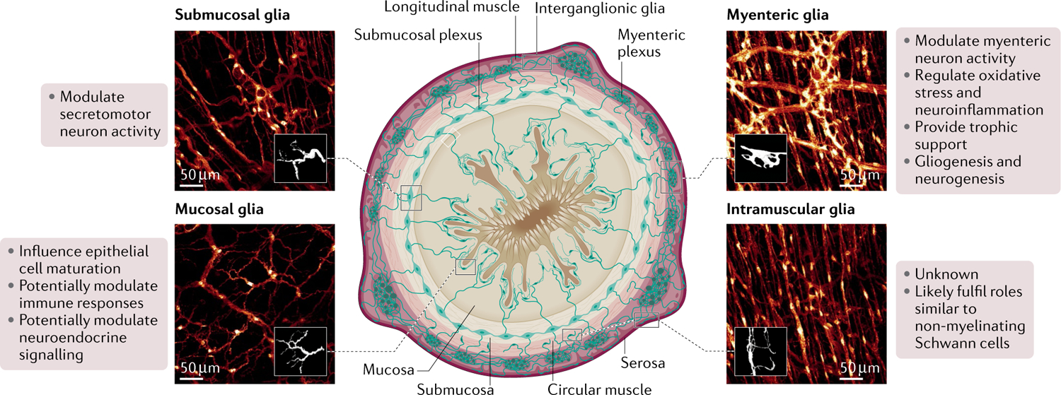
Fig. 1 |. Main populations of enteric glia and their known physiological functions.

Local subpopulations of glia are defined based on their morphology, anatomical location throughout the intestinal wall, and localization either within or outside of enteric ganglia. Based on these criteria, at least six main types of enteric glia are identified in any given region of the intestine: intraganglionic glia, including glia associated with neuronal cell bodies in the myenteric and submucosal plexuses (myenteric glia or Type-IMP and submucosal glia or Type-ISMP, on the top right and left, respectively); interganglionic glia, located within nerve fibre bundles connecting myenteric ganglia (Type-II); extraganglionic glia, including glia associated with nerve fibres at the myenteric and submucosal plexus but outside the ganglia (Type-IIIMP/SMP) and in the intestinal mucosa (mucosal glia or Type-IIImucosa, bottom left); and glia associated with nerve fibres in the circular and longitudinal muscle layers (intramuscular glia or Type-IV, bottom right). Known functions of each subtype are listed beside each representative image.
