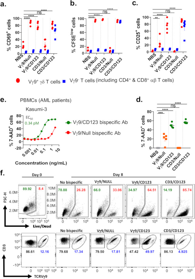
Fig. 4. Vγ9/CD123 bispecific antibody mediates selective activation, proliferation, and effector functions of Vγ9+ γδ T cells among whole PBMCs.
CFSE-labelled whole PBMCs from healthy individuals were cultured with Kasumi-3 cells in the presence or absence of the indicated bispecific antibodies at a concentration of 3 ng/mL. Scatter plot graphs mirror the mean (±SEM) frequency of Vγ9+ γδ T cells and Vγ9 TCR-depleted T cells that were positive for surface expression of CD69 (a), CFSE dilution (proliferation profile, b), CD25 (c), and the ability to eliminate exogenously spiked-in Kasumi-3 cells (cytotoxicity, d). The red circle and blue squares represent Vγ9+ (γδ) T cells and Vγ9 TCR-depleted T cells, respectively. Each dot represents data from an individual donor. (e) AML patient PBMCs cultured in the presence of Zol+IL-2+IL-15 for 14 days (effectors) were co-cultured with Kasumi-3 (target) cells at indicated concentrations of Vγ9/CD123 or Vγ9/Null arm control bispecific antibodies for 16–24 h. Target cell lysis was assessed by 7-AAD+ staining on flow cytometry. Cytotoxicity values represented in (e) are after subtracting cell death from no bispecific (NBS) antibody controls. Mean EC50 value of Vγ9/CD123 (green) bispecific mediated lysis of Kasumi-3 (0.34 pM) was from two patients. The control Vγ9/Null (red) bispecific antibodies showed non-target specific cytotoxicity (e). f Whole PBMCs from AML patients were cultured either in the presence or absence of indicated bispecific antibodies for 8 days. The number in the representative FACS dot plots refers to live and dead cells among AML blasts (f, upper row) and the frequency of Vγ9− CD3+ and Vγ9+ CD3+ cells among total CD3+ cells (f, lower row) on day 0 and 8 of the culture period. Representative data are means of values derived from two AML patients. The p values were calculated with a one-way ANOVA and Dunnett’s multiple comparison test. (*p < 0.05, **p < 0.01, ***p < 0.001, ****p < 0.0001, and ns suggests p > 0.05). NBS no bispecific antibody.

