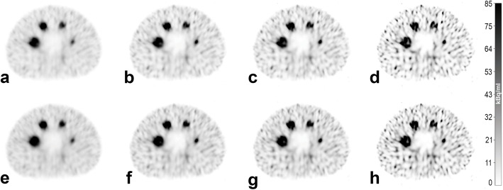
Fig. 3.
SPECT images reconstructed from data acquired with the clinical acquisition protocol. Reconstruction was performed with 2i/10s (a and e), 4i/10s (b and f), 5i/15s (c and g) and 24i/10s (d and h). Scatter correction was performed with a–d SCF = 1.10 and e–h SCF = 0.41. SPECT images (a and e) were reconstructed with postfiltering and represent the subjectively preferred image quality for reading. a represents our institutional standard for diagnostic imaging and reconstruction. All SPECT images have an identical window level and width

