Fig. 1.
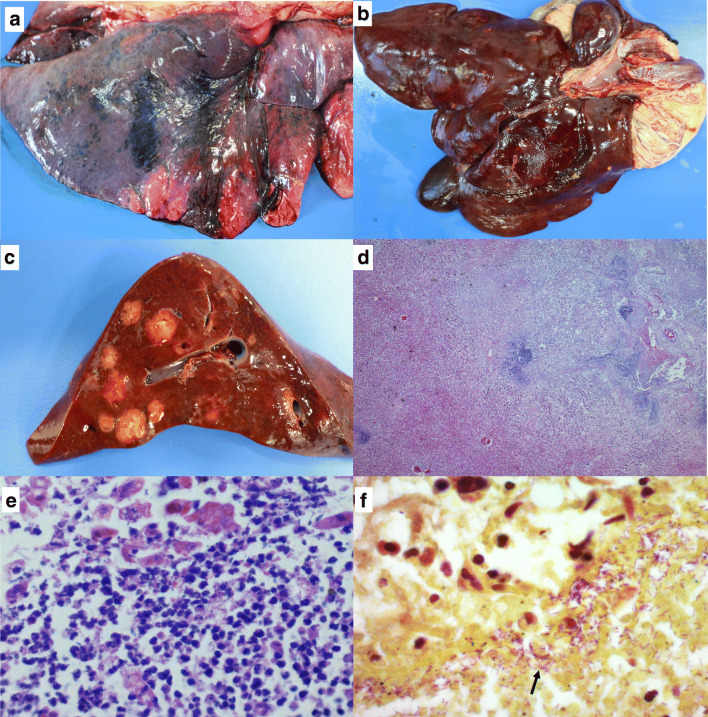
Sepsis in cougar (Puma concolor) associated with Chromobacterium violaceum: a Lung with an intensely dark red pleural surface associated with acute hemorrhage and congestion; b capsular surface of the liver with irregular nodules, multifocal to coalescent, yellowish, 0.5 to 2 cm; c cut surface of the liver with homogeneously yellowish, friable, well-demarcated nodules and with irregularly apparent capsule; d photomicrograph of the liver section with intense disorganization of the lobular structure with liquefactive hepatocellular necrosis and multifocal, random and extensive disappearance of parenchyma replaced by an intense suppurative infiltrate. H&E stain, ×2.5; e amplification of the anterior image showing an intense infiltration of neutrophils. H&E stain, ×63; and f section of the liver with marked pinkish purple bacillary structures (arrows). Brown-Hopps (Gram) stain, ×100

