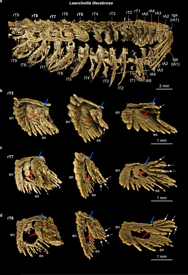
Fig. 1. Computed tomographic images of YKLP 11424 showing selected exite-bearing appendages of Leanchoilia illecebrosa.
a Ventral side of the animal. b–d Digitally dissected trunk appendages 5, 7, and 8 from the right side of the animal (rT5, rT7, rT8). Each appendage is shown at three different angles to demonstrate the endopodite (en), the exopodite (ex) and the exite consisting of one basal flap (red arrow) and several additional ones (white arrows). Blue arrows point to the attachment of the exite. Individual scale bars provided. An, head appendage n; l, left; r, right; ga, great appendage; Tn, trunk appendage n. Dissections of all appendages are available in Supplementary Figs. 2–4.

