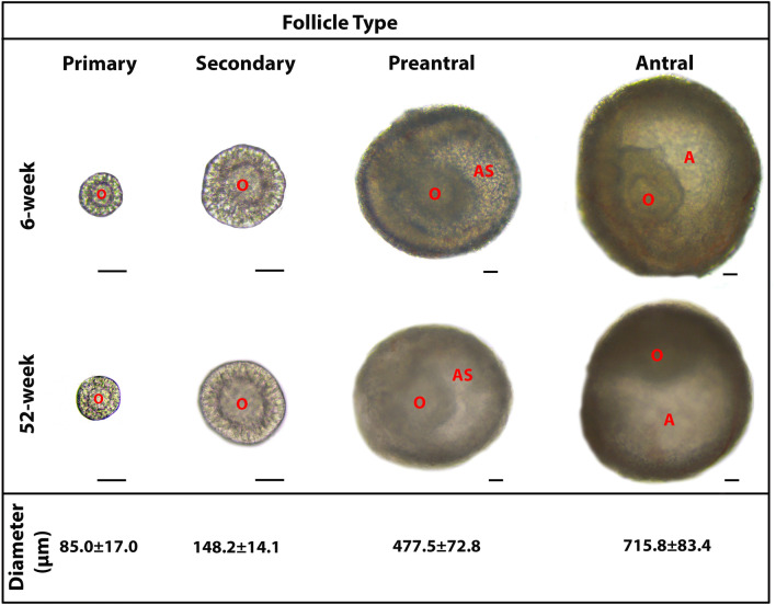
Figure 1.
The representative micrographs of the follicles from primary to antral stages, obtained from adult (6-week-old) and aged (52-week-old) mouse ovaries. The micrographs of the follicles were captured under a light microscope (Zeiss, Oberkochen, Germany). All the follicles displayed their characteristic features either in the adult or aged groups. Morphologically normal follicles at similar diameters were used in the subsequent experiments. The average diameter of each follicle type is provided below its own micrograph as mean ± standard deviation (SD). It should be noted that the micrographs of the primary and secondary follicles were captured under an original magnification of 200 ×, whereas those of the preantral and antral follicles were captured at an original magnification of 100 ×. Scale bars are equal to 50 µm. A antrum, AS antral space, O oocyte.

