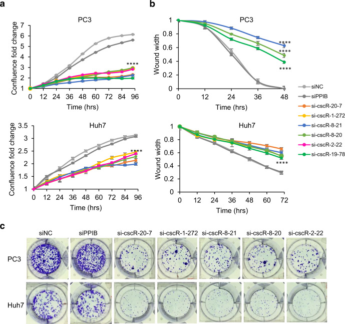
Fig. 4. Biological relevance of the cscRNAs in cancer cells.
a Cell proliferation curves of PC3 and Huh7 cells upon siRNA-mediated knockdown of the cscRNAs. The y-axis shows the cell confluence fold-change in relative to the confluence at time 0. The error bars represent the ±SD of three biological replicates. ****p = 0.0001 from two-sided t-test. Source data are provided as a Source data file. b Wound-healing assays showing the cell migration rates of PC3 and Huh7 upon siRNA-mediated knockdown of the cscRNAs. The y-axis shows the wound width at different time points in relative to the wound width at time 0. The error bars represent the ±SD of three biological replicates. ****p = 0.0001 from two-sided t-test. The representative images of the wounds are shown in Fig. S10. Source data are provided as a Source data file. c Formation of PC3 and Huh7 cell colonies upon siRNA-mediated knockdown of the cscRNAs.

