Fig. 1.
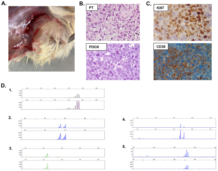
Characterization of EMM PDOX. (A) Macroscopic appearance of PDOX. (B) H&E staining of the patient's extramedullary lesion (PT, upper panel) and xenografted tumor (PDOX, lower panel), showing histological similarities between the original tumor sample and the originated EMM PDOX (at 400× magnification). (C) EMM PDOX cells showed a Ki67 positivity of 90% and CD38 expression, as observed in the patient's precursor tumor. (D) Paired microsatellite genotyping of PDOX and the patient's lesion from which it was derived. The genetic match between the generated PDOX and the patient's tumor was assessed by microsatellite genotyping. The microsatellites used for tumor genotyping were (1) D5S299, (2) D5S346, (3) D3S1612, (4) D5S82 and (5) D3S3564. For each microsatellite, the upper panel corresponds to the patient's biopsy and the lower panel corresponds to PDOX.

