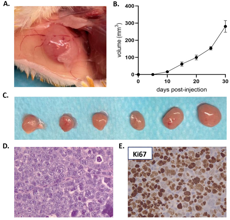
Fig. 2.

Derived EMM cell line generated plasmocytomas when re-implanted in NSG mice. (A) Representative image of a plasmocytoma derived from EMM cell line by intradermal injection of 1×10⁶ cells soaked in Matrigel in the flank of an NSG mouse. (B) Tumor growth rate of plasmocytomas generated after the intradermic injection of 1×10⁶ cells of the EMM cell line (n=6). (C) Tumors reached a mean volume of 280±34 mm3 at 30 days post-injection. (D) H&E staining of the cell line-derived plasmocytomas showing histological similarities with the PDOX and the patient's biopsy (at 400× magnification). (E) Cell line-derived plasmocytomas showed a high Ki67 expression as observed in the EMM PDOX (at 400× magnification). Data are mean±s.e.m.
