
Fig. 3.
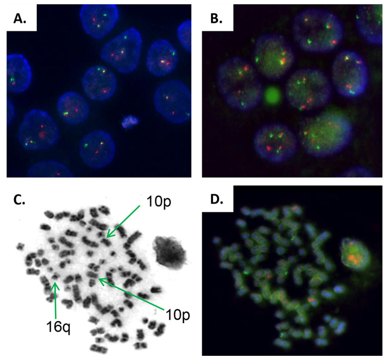
Fluorescence in situ hybridization analysis of the immunoglobulin heavy chain gene locus (IGH). (A) IGH rearrangement was observed in only one chromosome at diagnosis before the occurrence of extramedullary disease (Break Apart Probe). (B) FISH analysis of t(14;16) IGH-MAF rearrangement of EMM PDOX showing the translocation in the two chromosomes. (C,D) Micrograph of G-band metaphase chromosomes of the PDOX (C) and FISH (D) analysis of IGH rearrangements, identifying chromosomes 10 (region p) and 16 (region q) implicated in IGH t(14;16)(q32;q23) and t(10;14)(p?11-12;q32) translocations. The later translocation was not detected at MM diagnosis. All images are at 1000× magnification.