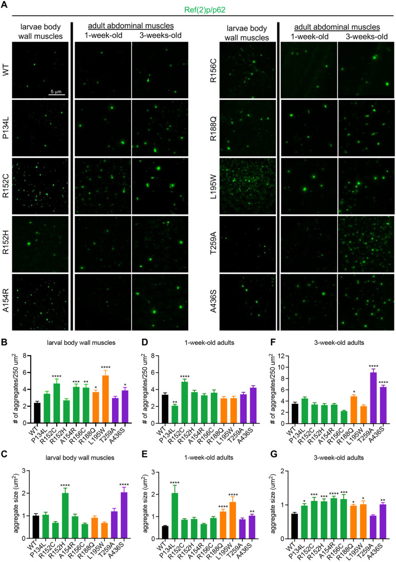
Fig. 5.
Proteostasis analysis in VCP disease models. (A) Representative images of p62 staining in larval body wall muscles and adult abdominal muscles at 1 week and 3 weeks of age in the indicated genotypes. (B,C) Quantification of the number (B) and size (C) of aggregates observed in larval body wall muscles. n=4 independent animals. (D,E) Quantification of the number (D) and size (E) of aggregates observed in 1-week-old adult abdominal muscles for the indicated genotypes. (F,G) Quantification of the number (D) and size (E) of aggregates observed in 3-week-old adult abdominal muscles for the indicated genotypes. Data are mean±s.e.m. For number of aggregates, n>50 muscles from four independent animals. For aggregate size, n>100 aggregates from four independent animals. *P<0.05, **P<0.01, ***P<0.001, ****P<0.0001 (one-way ANOVA with Dunnett's multiple comparisons). WT, wild type.

