Figure 1:

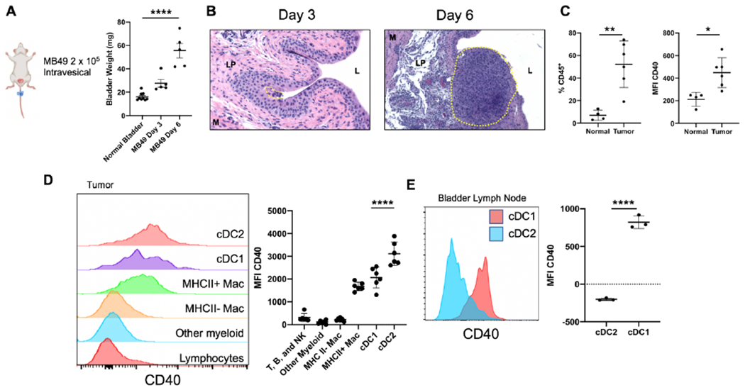
CD40 is expressed on dendritic cells within the bladder tumor microenvironment.
A, Schematic of orthotopic tumor model with bladder weights (mg) at days 3 and 6 post intravesical tumor implantation. B, Histology (H & E) of bladders from days 3 and 6 post tumor implantation. Tumor area is outlined by the yellow dotted line. The bladder lumen (L), lamina propria (LP), and muscle layer (M) are denoted by their respective letters. C-D, Flow cytometry of MB49 bladder tumors. (C) percentage of CD45+ immune cells and mean fluorescent intensity (MFI) of CD40 on CD45+ immune cells in normal (n = 4 independent animals) and tumor-bearing bladders (n = 6 independent animals). (D) histograms and quantification of CD40 expression across immune cell types in the tumor-bearing bladder. E, Flow cytometry histogram and quantification of CD40 expression in bladder-draining lymph nodes across DC subtypes. cDC1 are defined as CD11c+ MHCII+ Xcr1+. cDC2 are defined as CD11c+ MHCII+ Sirpα+. For A and D, One-way ANOVA with Tukey’s test was used to calculate statistical significance, *** p < 0.001, **** p < 0.0001. For C and E, Two-tailed unpaired t-test was used to calculate statistical significance, * p < 0.05, ** p < 0.01, **** p < 0.0001. All data are plotted as mean ± s.d. Data are representative of two independent replicates.
