Fig. 5.
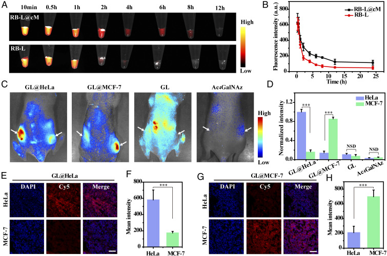
In vivo selective fluorescence imaging of tumor-associated glycans using GL@cM. (A) Fluorescence images of blood samples from mice injected with RB-L and RB-L@cM. (B) In vivo pharmacokinetic curves of RB-L and RB-L@cM as quantified by fluorescence spectroscopy (n = 3). (C) In vivo fluorescence visualization of mice with tumors administrated with GL@HeLa (60 mg ⋅ kg−1), GL@MCF-7 (60 mg ⋅ kg−1), GL (60 mg ⋅ kg−1), and Ac4GalNAz (60 mg ⋅ kg−1) on 4 consecutive days. DBCO-Cy5 was intravenously injected into the mice on day 5. (D) Quantitative analysis of the normalized fluorescence intensity. Data are presented as mean ± s.d. (n = 3). (E and F) Fluorescence photographs and intensity analysis of tumor sections from mice injected with GL@HeLa. (G and H) Fluorescence photographs and intensity analysis of tumor sections from mice injected with GL@MCF-7. Cell nucleus was stained with DAPI. Fluorescence intensity of confocal microscopy images was analyzed by using Nikon Eclipse Analysis software. Data were presented as mean intensity (n = 10). (Scale bars: 50 µm.) Asterisks indicate significant differences (NSD: no significant difference, ***P < 0.001).

