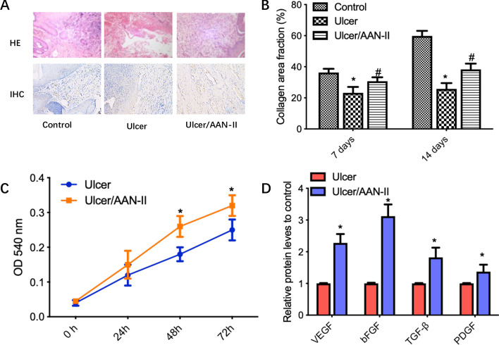
Fig. 2.
AAN-II promotes fibroblast proliferation and secretion of proangiogenic factors. A Hematoxylin and eosin (HE) staining was used to determine the skin pathological changes and immunohistochemical staining with anti-vimentin antibody was performed in the three groups. Representative fields are shown (×200). B The collagen area fraction (%) was calculated on days 7 and 14 and values are expressed as means ± SEM. *p < 0.05 compared with the control group. #p < 0.05 compared with the ulcer group. C CCK-8 assay was used to determine the proliferation ability of rabbit skin fibroblast cells with or without AAN-II treatment. Values are expressed as means ± SEM. *p < 0.05 compared with the control group. D The relative protein levels of the proangiogenic factors VEGF, bFGF, TGF-β and PDGF were measured using ELISA assays. Values are expressed as means ± SEM. *p < 0.05 compared with the control group

