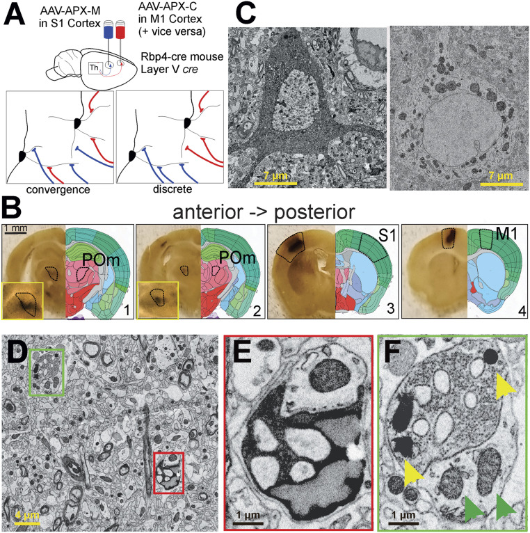
Fig. 1.
Summary of experimental strategy. (A) We injected two types of AAV-expressing cre-dependent APX into the primary S1 or primary M1 cortices of two Rbp4-cre transgenic mice; this strategy limited expression of the label to layer 5 cells and in particular to layer 5 corticothalamic terminals in POm. In one mouse, as shown, the APX-C label was placed in M1 and the APX-M label in S1; in the other mouse (not shown) the APX-C label was placed in S1 and the APX-M label in M1. (B) DAB reactions in serial vibratome slices revealed APX expression in layer 5 cells of the cortical areas targeted as well as labeled layer 5 terminals in POm. (C) Electron micrographs of layer 5 neuronal cell bodies in S1 and M1 cortices show clearly distinguishable labeling when APX-C labels the cytoplasm (Left) and APX-M labels the mitochondria (Right). (D–F) Electron micrographs show two terminals near one another, one from layer 5 of M1 labeled with APX-C (red box in D and E) and the other from S1 labeled with APX-M (green box in D and F). (Insets) Higher resolution images of the synapses, and in F, yellow arrowheads point to synapses with APX-labeled mitochondria, and green arrowheads point to unlabeled mitochondria in other neurons.

