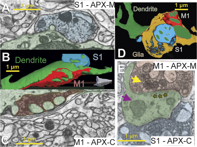
Fig. 3.
Convergence of layer 5 M1 and S1 inputs onto individual POm dendrites. We found M1 and S1 synapses on the same POm dendrites in both animals. Shown are individual electron micrographs (A, C, and E) corresponding to three-dimensional reconstructions (B and D) of individual POm relay cell dendrites (green) from two mice. Both dendrites are postsynaptic to large layer 5 terminals from M1 (red) and S1 (blue) labeled with a different APX-targeted subcellular compartment in each animal. In A through C, the animal was injected with APX-C in M1 and APX-M in S1, and for D and E, the labels were switched, APX-C in S1 and APX-M in M1. Each input and the dendrite are shaded in the electron micrographs (A, C, and E) corresponding to their colors in the reconstructions (B and D). Synaptic vesicles can be clearly seen (A, C, and E), and a clear postsynaptic density is shown in E (asterisks). Finally, examples of mitochondria labeled with APX (yellow arrow) as compared to unlabeled mitochondria in the postsynaptic dendrite (purple arrow) are shown in E.

