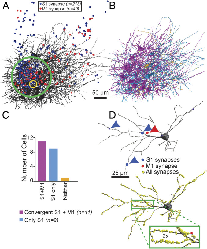
Fig. 4.
Convergence of M1 and S1 inputs on single thalamic neurons in POm is common. (A) In the mouse with APX-C labeling from M1 and APX-M labeling from S1, we first identified regions (green circle) in the electron microscopic datasets with the highest concentrations of labeled S1 (blue circles) and M1 (red circles) terminals. The yellow circle indicates the region in which detailed measure of all synapses identified to determine synaptic density. (B) In that volume, we reconstructed 2.74 mm of the full dendritic arbor of all 21 neurons and identified all S1- and M1-labeled inputs on each neuron. (C) We found that 20 of the 21 neurons had at least a single input from S1 (cyan neurons) and that 10 of these showed a second input from the M1 cortex (purple). We found no examples of innervation of these POm neurons by M1 and not S1 and only one example of a neuron that received neither labeled input (orange neuron in B and C). (D) For subset of four neurons with convergent input, we determined the location and size of all synapses labeled or unlabeled (yellow spheres). An example is shown, and unlabeled synapses are indicated as yellow circles.

