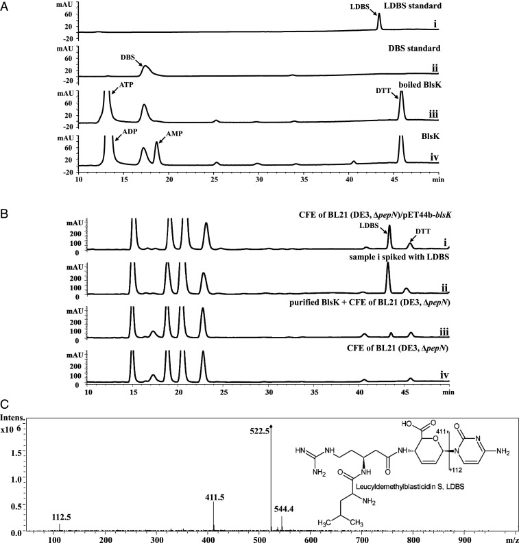
Fig. 3.
Functional analysis of BlsK in vitro. (A) Assay with purified BlsK: (i) the LDBS standard; (ii) DBS standard; (iii) boiled BlsK as a negative control; and (iv) assay with 1 mM ATP, 10 mM leucine, and 250 μM DBS. (B) Assay of BlsK with CFE: (i) CFE of BL21 (DE3, ΔpepN)/pET44b-blsK with DBS; (ii) sample i spiked with LDBS standard; (iii) purified BlsK adding CFE of BL21 (DE3, ΔpepN) with DBS, and (iv) CFE of BL21 (DE3, ΔpepN) with DBS. In total, 1 mM ATP and 5 mM leucine were added in all groups. (C) MS/MS spectrum of the product of BlsK-catalyzed reaction (sample [B] 1): chemical structure of LDBS with the key fragmentation pattern shown. The reaction conditions were pH 8.0 and 37 °C for overnight.

