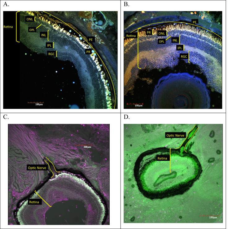
Figure 2. Ocular sections of adult E. nana (A, C) and E. sosorum (B, D).
Using a laser scanning confocal microscope to detect autofluorescence from the sections, montages of images were produced using digitally applied pseudocolors to provide contrast. Ocular sections from adult E. nana (A and C) and adult E. sosorum (B and D) and associated retinal layers and optic nerve (C and D). Layers include pigment epithelium (PE), photoreceptor layer (PR), outer nuclear layer (ONL), outer plexiform layer (OPL), inner nuclear layer (INL), inner plexiform layer (IPL), and retinal ganglion cell layer (RGCL).

