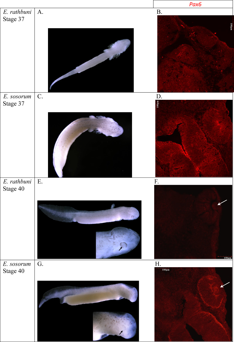
Figure 8. E. rathbuni and E. sosorum embryos at two stages of development and sections illustrating Pax6 labeling.
The top two rows (A–D) of images show embryos at stage 37 (A & C) with their respective histological sections (B & D); the bottom two rows (E–H) of images show embryos at stage 40 (E & G) with black arrows indicating eye development. Histological sections for stage 40 (F & H) show labeling for Pax6 and lens development (white arrows). The image of a histological section through the anterior part of a stage 37 E. rathbuni embryo shows tissue positive for Pax6 labeling, where aggregates (not labeling; B) of secondary antibody are also evident (negative controls: File S2).

