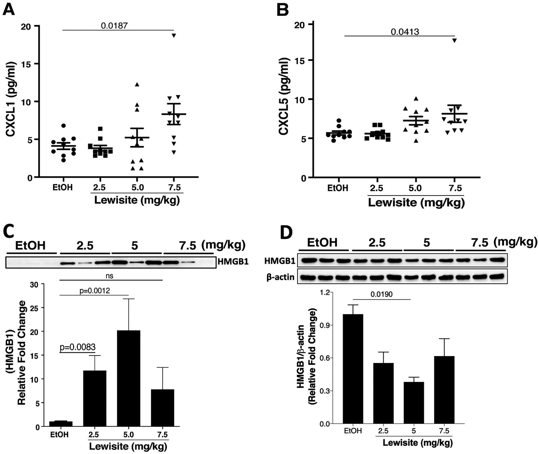
Figure 5. Cutaneous Lewisite exposure increases CXCL1, CXCL5 and HMGB1 in BALF.

Cutaneous Lewisite exposures were performed at different doses as described above and mice were sacrificed 24 hours later. BALF was collected as described in the Methods. Concentrations of (A) CXCL1 and (B) CXCL5 in BALF of animals exposed to either EtOH (ethanol: control) or increasing doses of Lewisite are shown. Values are expressed as mean±SEM. Immunoblotting of HMGB1 was performed on (C) BALF and (D) lung tissue samples from control and Lewisite treated animals. The top panels demonstrate representative HMGB1 immunoblots. Densitometric analysis of blots are shown as a graph in the lower panel. Densitometric analysis of HMGB1 immunoblot for lung tissue lysates is plotted, relative to the expression of β-actin. Values are expressed as mean±SEM.
