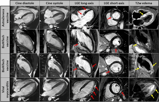
Fig. 2.
CMR images of patients showing patterns of myocardial damage after COVID-19 vaccination. Cardiovascular magnetic resonance (CMR) images including cine images at diastole and systole (first two columns), late-gadolinium-enhancement (LGE) images in long-axis and short-axis views (third and fourth column) as well as T2-weighted edema images in long-axis views (fifth column). The individual CMR-based cardiac phenotype of (a) patient 1 after 1st dose of AstraZeneca vaccine (upper panel), (b) patient 2 after 1st dose of Pfizer–BioNTech vaccine (second panel) and (c) patient 3 after 2nd dose of Pfizer–BioNTech vaccine (third panel) are illustrated. For comparison, the bottom panel shows CMR findings of a young patient that suffered from viral myocarditis

