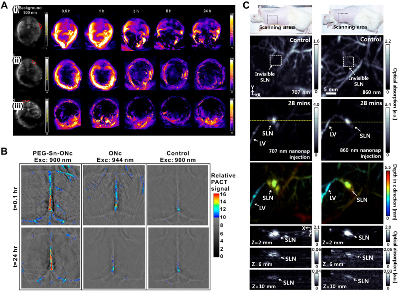
FIG. 3.
Contrast-enhanced PA images. (a) In vivo PA signals at various time points after tail-vein administration of (i) PcS4, (ii) ZnPcS4, and (iii) AlPcS4. Reprinted with permission from Attia et al., Biomed. Opt. Express 6(2), 591 (2015). Copyright 2015 The Optical Society. (b) Noninvasive photoacoustic images of brain blood vessels of mice administered with PEG-Sn-ONc or ONc at the indicated time points following intravenous injection. Reprinted with permission from Huang et al., Bioconjugate Chem. 27(7), 1574 (2016). Copyright 2016 American Chemical Society. (C) In vivo PA imaging of rat's SLNs with injection of 707 nm (left column) and 860 nm (right column) nanonaps. Reprinted with permission from Lee et al., “Dual-color photoacoustic lymph node imaging using nanoformulated naphthalocyanines,” Biomaterials 73, 142–148. Copyright 2015 Elsevier. PA, photoacoustic; SLN, sentinel lymph node; LV, lymphatic vessel.

