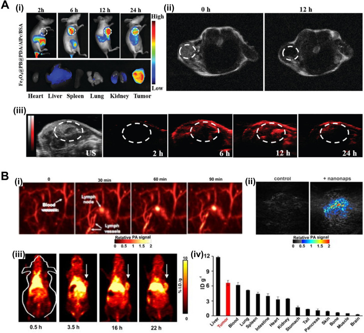
FIG. 4.
Pc/Ncs for multimodal imaging. (a) (i) In vivo FL images after i.v. injection of Fe3O4@PB@PDA/AlPc/BSA in tumor-bearing mice at 2 h, 6 h, 12 h, 24 h and ex vivo FL images of AlPc in major organs induced with 660 nm laser irradiation after i.v. injection for 24 h. (ii) MR signals in the tumor before and 12 h after i.v. injection with Fe3O4@PB@PDA/AlPc/BSA. (iii) US and PA images of tumor-bearing mice after i.v. injection with Fe3O4@PB@PDA/AlPc/BSA taken different time points. Republished with permission from Wang et al., J. Mater. Chem. B 6(16), 2460 (2018); permission conveyed through Copyright 2018 Royal Society of Chemistry, Clearance Center, Inc. (B) Nanonaps for PA and PET imaging. (i) PA lymph node imaging using nanonaps. (ii) PA imaging of subcutaneous 4T1 whole tumors in living BALB/c mice with or without i.v. administration of 2.6 mg nanonaps 24 h prior. (iii) Serial PET images of 4T1 subcutaneous breast tumors in BALB/c mice after i.v. injection. Arrows show tumor location. (iv) Biodistribution of 64Cu within nanonaps 24 hours post injection of nanonaps. Republished with permission from Zhang et al., Nanoscale 9(10), 3391 (2017); permission conveyed through Copyright 2017 Royal Society of Chemistry Clearance Center, Inc. FL, fluorescence; i.v., intravenous; MR, magnetic resonance; US, ultrasound; PA, photoacoustic; PET, positron emission tomography.

