Figure 2.

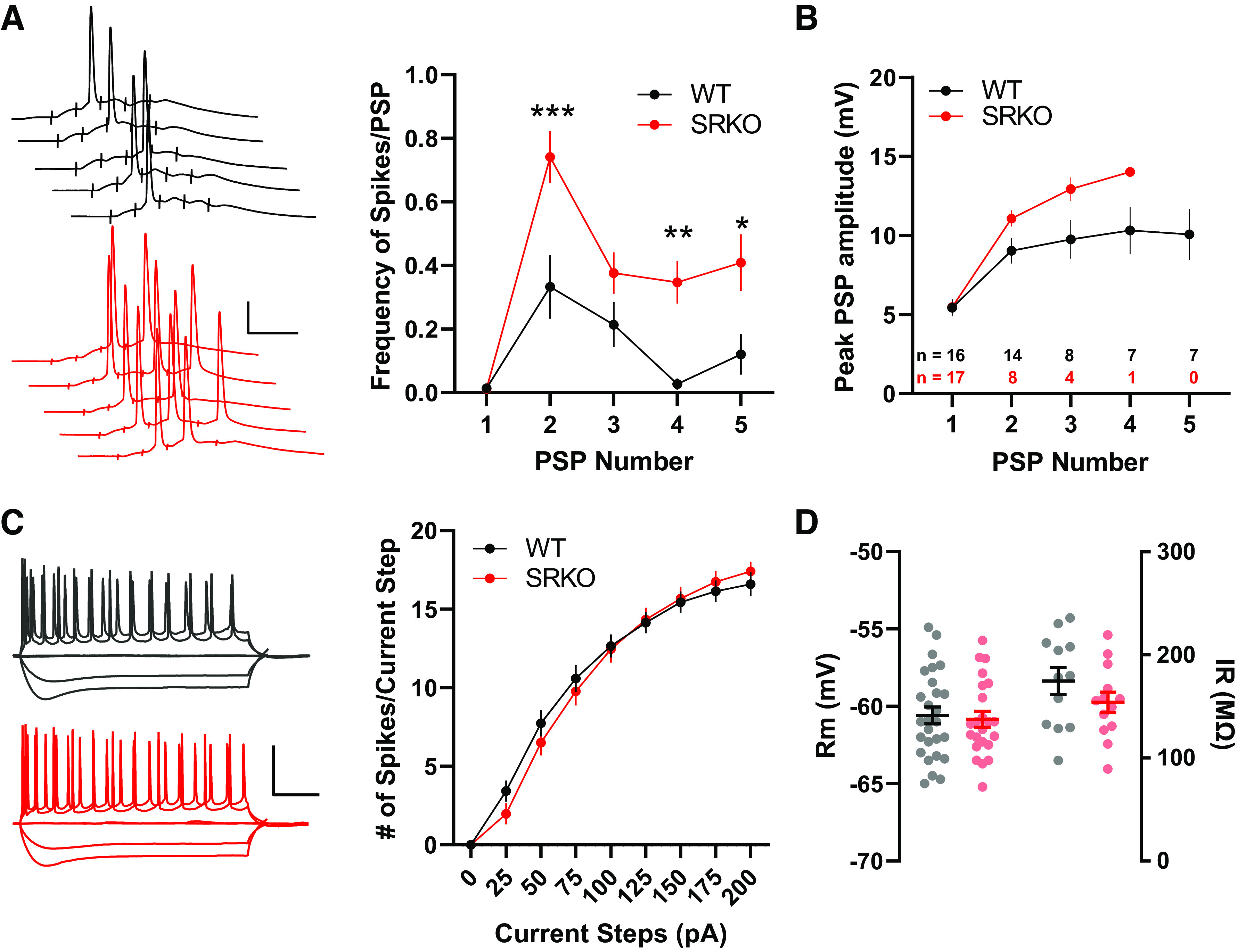
Increased synaptic excitability in SRKO mice. A: left, sample traces of APs/PSPs evoked by 5 pulses at 100 Hz Schaffer collateral fiber stimulation; scale bars: 25 mV, 20 ms. Right, short trains of synaptic stimulation leads to significantly more APs/PSP in SRKO compared with WT (PSP 1: P = 0.0001, two-way ANOVA, F(150) = 4.34, PSP 4: P = 0.004, two-way ANOVA, F(150) = 3.41, PSP 5: P = 0.013, two-way ANOVA, F(150) = 3.07; Bonferroni’s multiple comparisons test); WT: n = 15, SRKO: n = 17). B: temporal summation of PSPs measured from the 100 Hz stimulation in A until the first action potential for each cell (final n for each PSP shown in inset). C: left, sample traces for 0, −100, −200, +100, and +200 pA current steps; scale bars: 50 mV, 100 ms. Right, intrinsic excitability is unchanged in SRKO CA1 pyramidal neurons. Depolarization induced by somatic current injection elicits similar numbers of APs in WT and SRKO cells (P = 0.759, two-way ANOVA, F(8,207) = 0.6212; WT: n = 12, SRKO: n = 13) suggesting basal synaptic transmission is unaffected. D: summary graph of resting membrane potential (Rm) (right) and input resistance (IR) (left) showing no significance difference between WT and SRKO CA1 pyramidal cells. Data represent means ± SE. AP, action potential; PSP, postsynaptic potential; SRKO, serine racemase knockout; WT, wild-type. *P < 0.05, **P < 0.01, ***P < 0.001.
