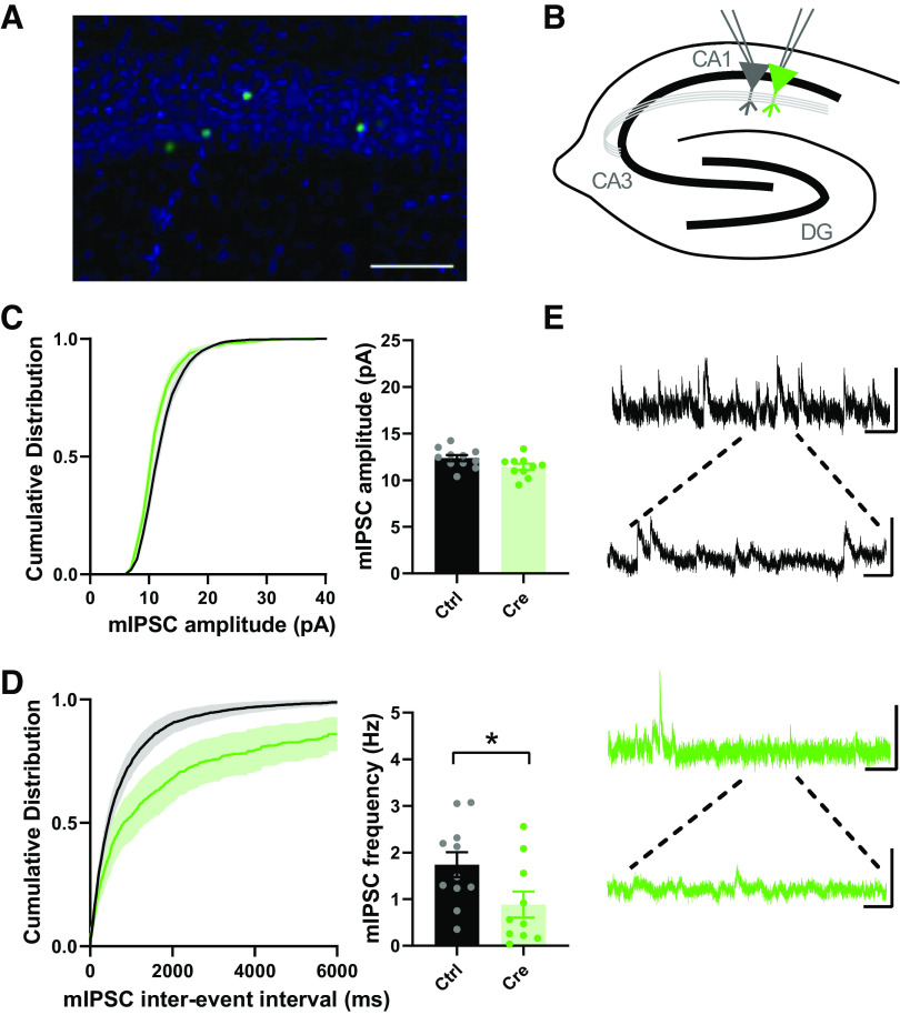
Figure 7.
Cell-autonomous reductions in spontaneous GABAergic synaptic transmission onto CA1 pyramidal cells following single-neuron SR deletion. A: representative image of the sparse transduction of CA1 pyramidal cells by AAV1-Cre:GFP counterstained by DAPI. Scale bar indicates 100 μm. B: schematic of the experimental setup. Whole cell mIPSC recordings were made from transduced (Cre+) and control CA1 pyramidal cells. C: cumulative probability and mean mIPSC amplitude. Although cumulative probability (KS test, P < 0.0001) of mIPSC amplitude was significantly changed between Cre and Cre+ neurons, the mean mIPSC amplitude from Cre+ neurons was not significantly different than those from control cells (WT: 12.37 ± 0.32 pA, n = 11; SRKO: 11.42 ± 0.34 pA, n = 10; P = 0.939). D: cumulative probability of interevent intervals and mean frequency of mIPSCs. Cumulative probability (KS test, P < 0.0001) and mean frequency from Cre+ neurons were significantly decreased compared to control cells (WT: 1.74 ± 0.27 Hz, n = 11; SRKO: 0.88 ± 0.28 Hz, n = 10; P = 0.039. E: sample mIPSC traces from control (black, top) and Cre+ (green, bottom) pyramidal neurons; scale bars: 25 pA and 0.5 s; inset, 25 pA and 100 ms. Data represent means ± SE. mIPSC, miniature inhibitory postsynaptic current; SRKO, serine racemase knockout; WT, wild type. *P < 0.05.

