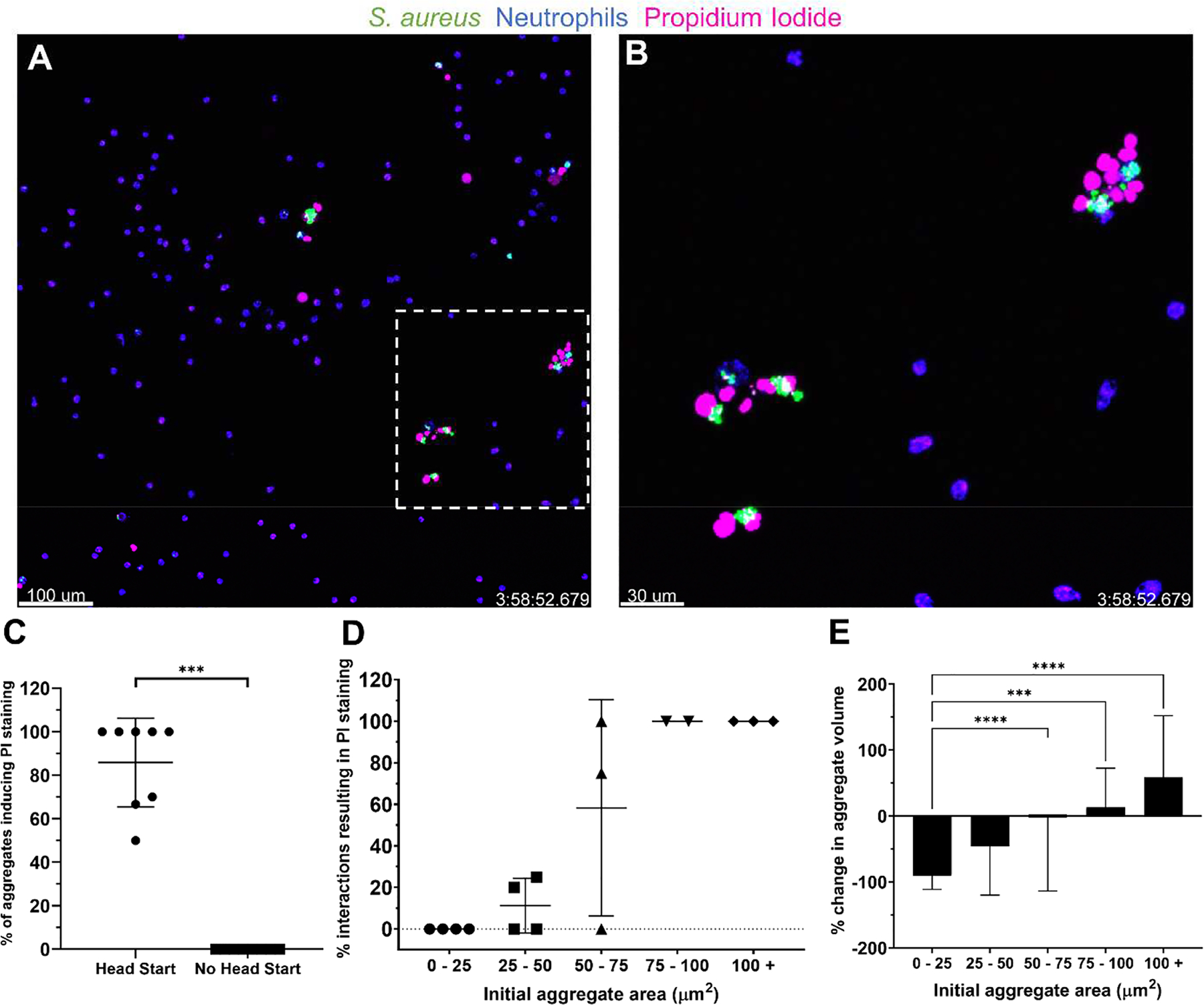
Figure 4:

Larger S. aureus aggregates cause neutrophil lysis and resist clearance in vitro. (A) S. aureus aggregates (green) were given 4 h to grow prior to the addition of neutrophils (blue). Representative images at t = 4 h show propidium iodide (PI) staining (pink) of neutrophils interacting with larger, persistent clusters of S. aureus. (B) Enlarged image of the inset in (A). (C) Percentage of S. aureus aggregates causing PI staining of at least one neutrophil. Data are mean ± SD (n=8 FOVs per group from 4 independent experiments. Mann-Whitney test ***p<.001). (D) Rates of PI staining of neutrophils as a function of the bacterial aggregate area at the beginning of the interaction. Each point represents the calculated average of all interactions within the size boundaries from a given experiment. The number of observed interactions per point ranged from n = 1–19 collected from 16 FOVs from 4 independent experiments. (E) Change in volume of an aggregate from the first frame in which a neutrophil phagocytoses it to the last frame that it is visible. Data are mean ± SD (Bins contain 6–58 measurements from 16 FOV from 4 independent experiments. Kruskal-Wallis test ***p<0.001, ****p<0.0001).
