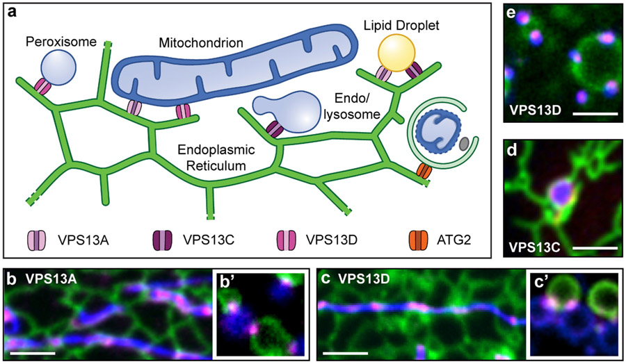
Fig. 2.
Mammalian VPS13 proteins localize at membrane contact sites. (a) Cartoon depicting the localization of VPS13A, VPS13C, VPS13D and ATG2 at contact sites between the ER and other organelles. Fluorescence panels (b to e) are snapshots from live cell imaging experiments showing the contacts indicated with the corresponding letter in the main panel. In each image the ER marker is in green, the specific VPS13 protein is in magenta and the tethered organelle is in blue: mitochondria in b and b′ and c and c′, an endolysosomes in d and peroxisomes in e. Panels b′, c′ and e are from cells exposed to hypotonic shock. Such treatment induces vesiculation of organelles [130] which remain connected by tethering proteins, allowing a better visualization of VPS13 localization at organelle contact sites. Micrographs of b, c, c′ and d were cropped from data published in [15,40]. Micrographs b′ and e are unpublished data (panel b′ from M. Leonzino and panel e from M. Leonzino and A. Guillén-Samander).

