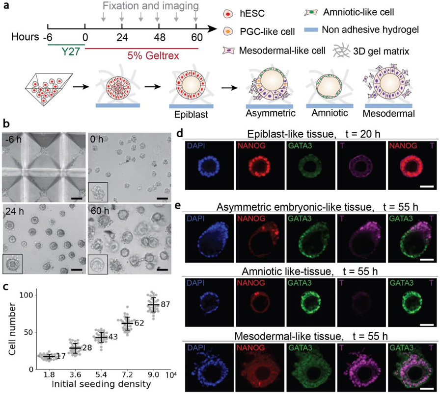
Figure 1. Guided self-organization of human pre-gastrulation embryonic-like tissues.
a, Schematics of generating and long-time culturing three types of tissues from hESCs, including asymmetric embryonic-like tissues, amniotic-like tissues, and mesodermal-like tissues. b, Representative bright-field images of cells in the pyramid well array and tissues in the 3D gel matrix at different time points as indicated. Scale bars, 100 μm. c, Number of cells inside a single pyramidal well as a function of the initial cell seeding density. d, Representative confocal images of the epiblast-like tissues at 20 h stained for DAPI, NANOG, GATA3 and T. Scale bar, 50 μm. e, Representative confocal images of the three types of embryonic-like tissues at 55 h stained for DAPI, NANOG, GATA3 and T as indicated. Scale bars, 50 μm.

