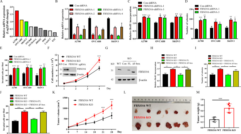
Fig. 2. Down-regulation of FBXO16 promotes ovarian cancer cell proliferation both in vitro and in vivo.
A The relative mRNA expression of FBXO16 in several ovarian cell lines from the Cancer Cell Line Encyclopedia (CCLE) database (https://portals.broadinstitute.org/ccle). B Ovarian cancer cells with or without FBXO16 silencing (FBXO16-shRNA1-4) were examined for FBXO16 mRNA expression. C Ovarian cancer cells with or without FBXO16 silencing were examined for BrdU cell proliferation. ***P < 0.001, **P < 0.01, *P < 0.05. D Cells in (C) were examined for colony formation. **P < 0.01,*P < 0.05. E Cells in (C) were examined for cell invasion. **P < 0.01. F FBXO16 knockout (KO) SKOV3 cells were generated by CRISPR assay and detected by western blot with indicated antibodies. The cell growth curve of control and FBXO16 KO cells was shown. ***P < 0.001. G FBXO16 KO SKOV3 cells infected with retrovirus coding FBXO16 wild-type (FBXO16 WT), or an F-box domain deleted mutant (FBXO16 ΔF). The cell lysates were subjected to immunoblot with indicated antibodies. H Cells in (G) were examined for BrdU cell proliferation. **P < 0.01, *P < 0.05. I Cells in (G) were examined for colony formation. **P < 0.01, *P < 0.05. J Cells in (G) were examined for cell invasion. ***P < 0.001, **P < 0.01. K Each nude mouse was subcutaneously injected with 1 × 10⁷ FBXO16 WT or FBXO16 KO SKOV3 cells for four weeks. Tumor growth was measured using a caliper at the indicated times after injection. n=6 for nude mice. ***P < 0.001. L Mice were sacrificed four weeks after transplantation. The tumors were then excised and photographed. M Tumor weights were measured after mice were sacrificed. ***P < 0.001.

