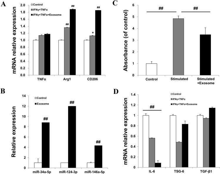
Fig. 3.
Exosomes derived from AMSCs induced an anti-inflammatory M2 phenotype in PBMCs. A Relative expression levels of TNFα, Arg1, and CD206 were determined by real-time PCR. PBMCs were co-cultured with IFNγ-and-TNFα-treated fibroblasts. Untreated fibroblasts were used as control. B Expression of miR-34a-5p, miR-124-3p, and miR-146a-5p was upregulated in exosomes compared to that in AMSCs. Exosomes derived from AMSCs exerted immunomodulatory effects and suppress inflammation. C The suppression of the proliferation of PHA-stimulated PBMCs was evaluated by using a proliferation assay kit. Unstimulated PBMCs were used as control. D The mRNA levels of IL-6, TSG-6, and TGF-β1 in PBMCs co-cultured with IFNγ-and-TNFα-stimulated fibroblasts were analyzed by real-time PCR. PBMCs co-cultured with unstimulated fibroblasts were used as control. The data are expressed as the mean ± SD of three independent experiments. *Significant difference from untreated control cells, p < 0.05. ##Significant difference from untreated control cells, p < 0.01

