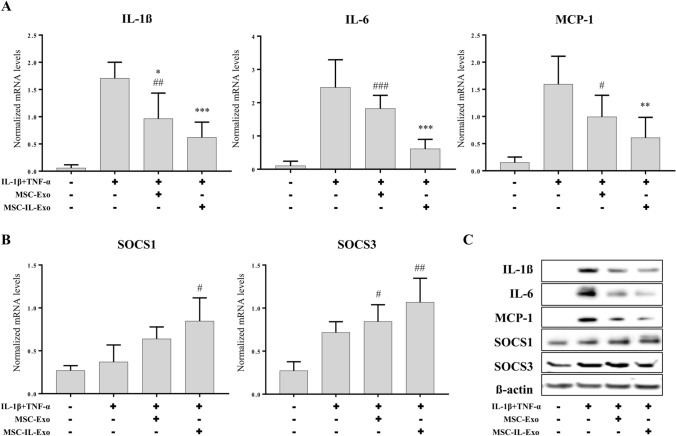
Fig. 2.
Anti-inflammatory activity of MSC-Exo and MSC-IL-Exo in SW982 cells. SW982 cells were treated with IL-1β (10 ng/mL) and TNF-α (25 ng/mL) for 24 h in the presence of 10 μg/mL MSC-Exo or MSC-IL-Exo. Expression of selected pro- or anti-inflammatory factors were analyzed via RT-qPCR and Western blot analyses. A mRNA levels of pro-inflammatory cytokines (IL-1β, IL-6, and MCP-1) normalized by that of GAPDH. B mRNA levels of anti-inflammatory proteins (SOCS1 and SOCS3) normalized by that of GAPDH. Data are presented as mean ± SD from three independent experiments (n = 3). #/*p < 0.05, ##/**p < 0.01, and ###/***p < 0.001 by one-way ANOVA for two exosome groups (MSC-Exo and MSC-IL-Exo) versus untreated control (#) or versus IL-1β and TFN-α groups (*). C Protein levels of the pro- or anti-inflammatory factors were examined via Western blot analysis. β-actin was used as an internal control

