Fig. 1.
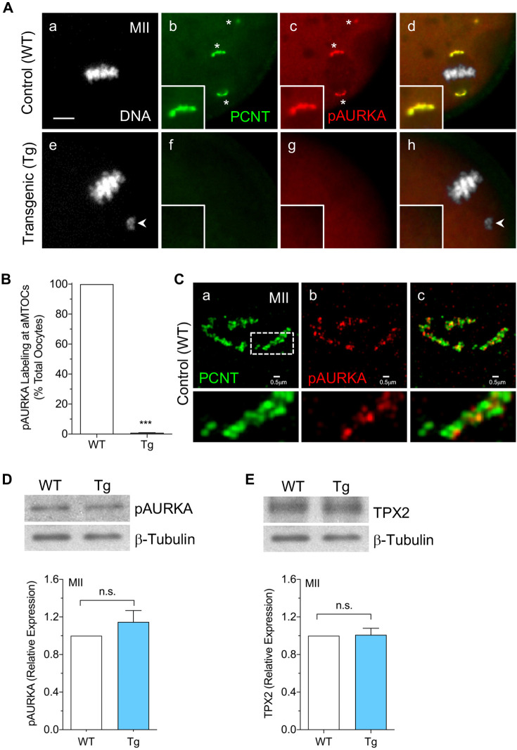
pAURKA localization to oocyte aMTOCs is dependent on PCNT. (A) Representative images of ovulated MII control (n=98) and PCNT-depleted (n=70) oocytes collected from WT and Tg mice, respectively, double labeled using anti-PCNT (green) and anti-pAURKA (red) antibodies at aMTOCs (*). DAPI-labeled DNA is shown in gray. Merged images are shown in panels d and h. Arrowheads indicate misaligned chromosomes. Insets show 2× magnification of the spindle pole. Scale bar: 5 µm. (B) Percentage of total oocytes with pAURKA labeling at aMTOCs. Data are presented as mean±s.e.m. of three experiments. (C) SR-SIM of PCNT (green) and pAURKA (red) colocalization at spindle pole aMTOCs in ovulated MII oocytes (n=7) from WT mice. Merged image is shown in panel c. Lower panels show 3× magnification of the region indicated by a dashed box. (D,E) Western blot analysis of (D) pAURKA and (E) TPX2 protein levels in ovulated MII oocytes (50 oocytes/lane) from WT and Tg mice. β-tubulin was used as an internal control, and total protein levels in WT oocytes are normalized to 1.0 for comparisons. Data are presented as mean±s.e.m. of three experiments. ***P<0.001, n.s., not significant (two-tailed, unpaired, Student's t-test).

