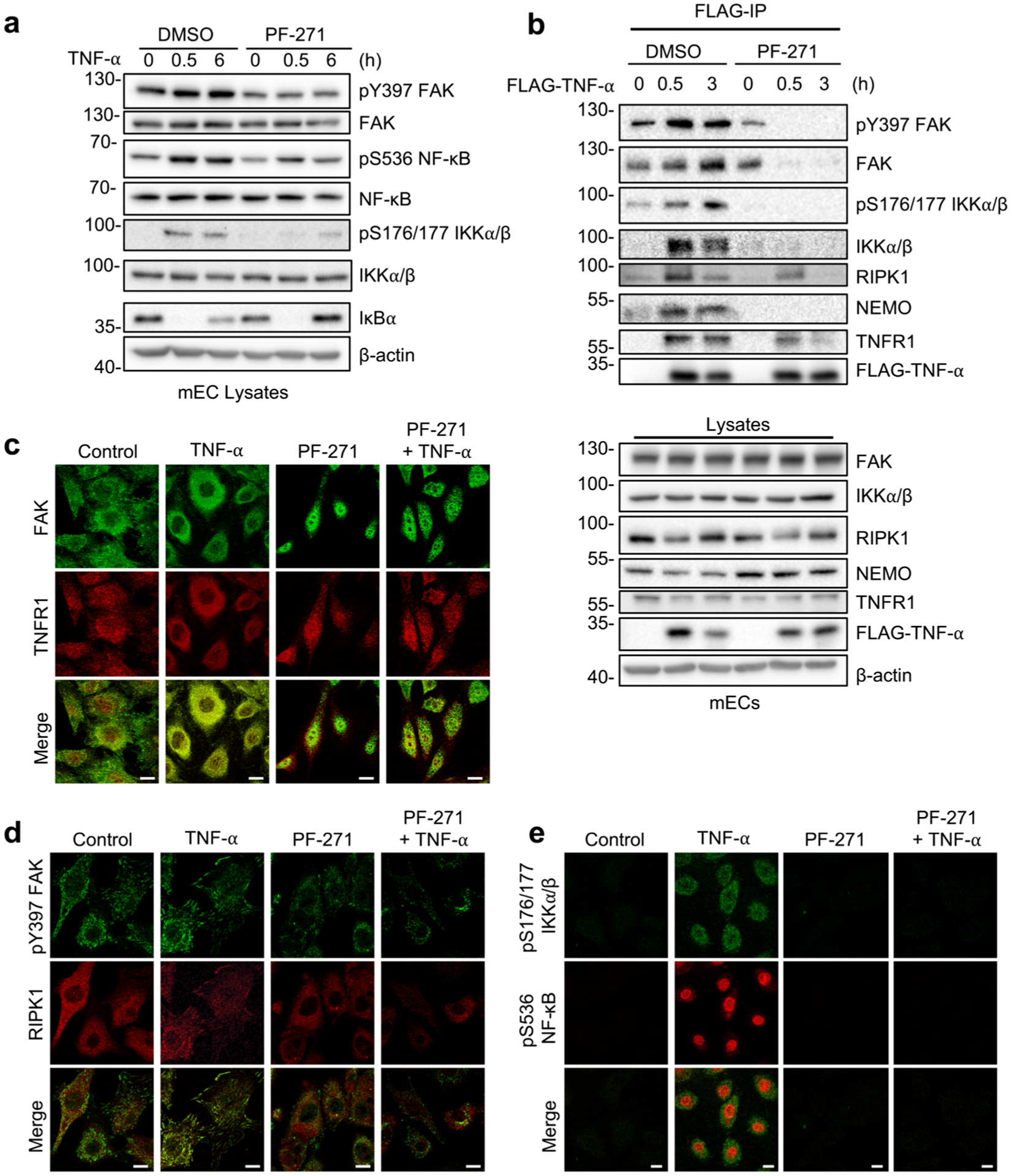
Fig. 3.

FAK inhibition disrupts RIPK1 and IKK recruitment to the TNF-α receptor. a, b mECs were treated for 1 h with DMSO or PF-271 (2.5 μmol/L) prior to TNF-α (10 ng/mL) stimulation for indicated times. a Western blot analysis of pY397 FAK, FAK, pS536 NF-κB, NF-κB, pS176/177 IKKα/β, IKKα/β, IκBα, and β-actin (n = 3). b Flag immunoprecipitation (IP) of FLAG-TNF-α stimulated mECs was performed. Western blot analysis for pY397 FAK, FAK, pS176/177 IKKα/β, IKKα/β, RIPK1, NEMO, TNFR1, FLAG-TNF-α, and β-actin in either FLAG-IP or lysates (n = 3). c–e mECs were treated for 1 h with DMSO or PF-271 (2.5 μmol/L) prior to TNF-α (10 ng/mL) stimulation for 3 h. c Immunostaining for FAK (green; rabbit) and TNFR1 (red; mouse) (n = 3). d Immunostaining for pY397 FAK (green; rabbit) and RIPK1 (red; mouse) (n = 3). e Immunostaining for pS176/177 IKKα/β (green; mouse) and pS536 NF-κB (red; rabbit) (n = 3). Scale bar, 20 μm.
