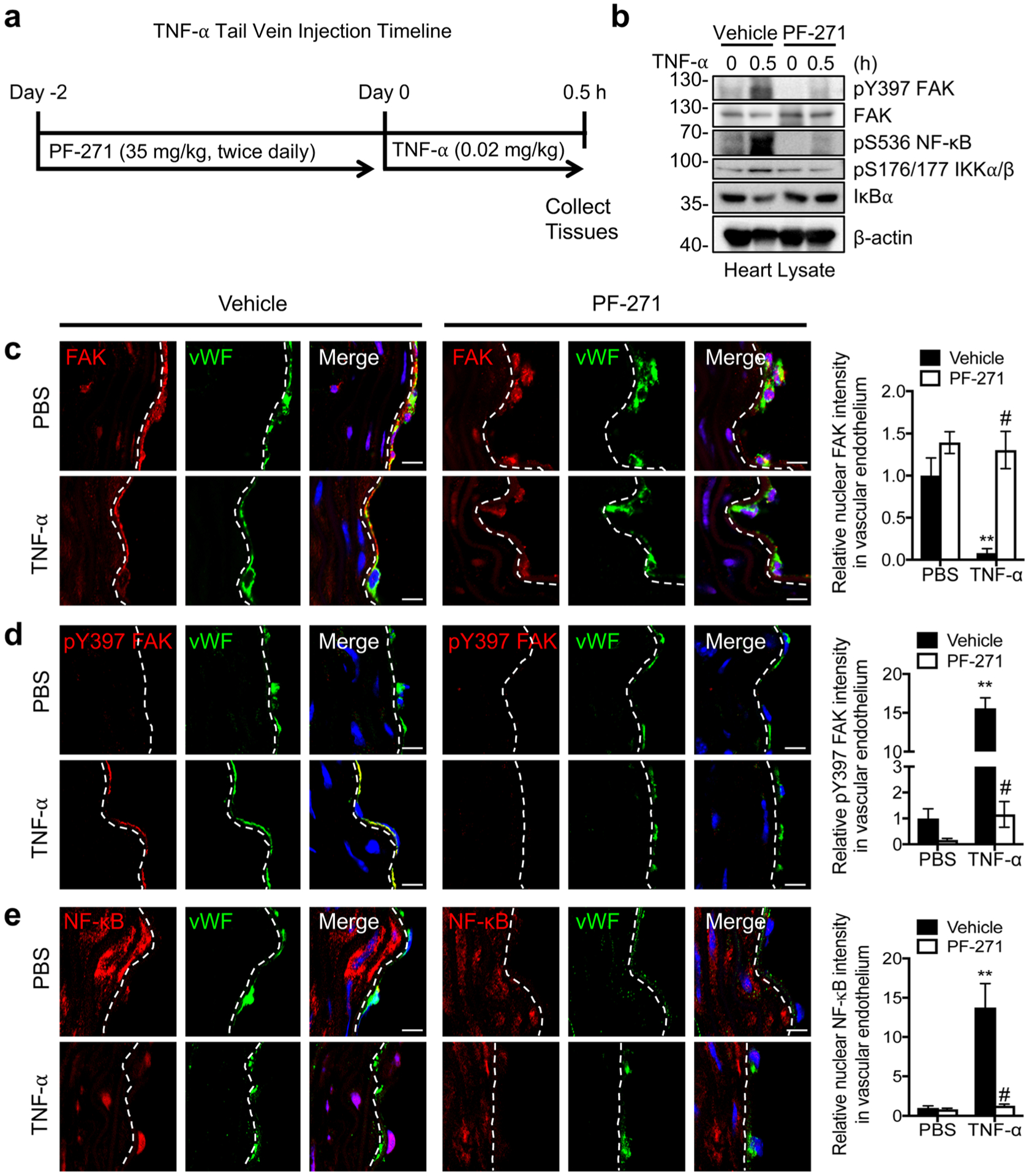
Fig. 4.

Immunostaining of mouse aortas showing that nuclear-localized FAK decreases TNF-α-induced NF-κB activation in mice. a Timeline for when C57BL/6 mice were treated with vehicle or PF-271 (35 mg/kg) before injection with PBS or mouse TNF-α (0.02 mg/kg) for 0.5 h. b Western blot analysis of heart lysates for pY397 FAK, FAK, pS536 NF-κB, pS176/177 IKKα/β, IκBα, and β-actin as loading control (n=4). Aorta sections were stained for FAK (red; mouse (c), pY397 FAK (red; rabbit) (d), or NF-κB (red; rabbit) (e). ECs were stained with vWF (green; rabbit or mouse) and nuclei with DAPI (blue). Relative fluorescence intensity in vWF-positive ECs was reported as mean ± SD (n = 4). Dashed lines, boundary between media and EC based on vWF staining. Scale bars, 10 μm. **P < 0.01 vs vehicle PBS; #P < 0.01 vs TNF-α.
